Khaos Group researchers participate in the creation of a tool based on Artificial Intelligence to improve the early detection of melanoma
A research team at the University of Malaga has developed a tool based on Artificial Intelligence and automatic image classification to improve the accuracy of detection of melanoma, a type of skin cancer.
The aim of the scientists is to understand how automation systems “think” in order to increase their efficiency and apply them in different fields of medicine. To this end, the experts propose a customized tool for each lesion detection program. This serves to improve the decision making of intelligent detectors used in the medical field, which ultimately help professionals to make diagnoses. In addition, it is a method that can be used with other similar screening programs, such as those focused on breast cancer.
As explained in their paper “Ensemble-based genetic algorithm explainer with automized image segmentation: A case study on melanoma detection dataset”, published in Computers in Biology and Medicine and available at: https://doi.org/10.1016/j.compbiomed.2023.106613, the experts have used an approach based on genetic algorithms (computational techniques inspired by biological evolution) and automatic image classification to analyze and detect patterns associated with melanoma in a set of photographs.

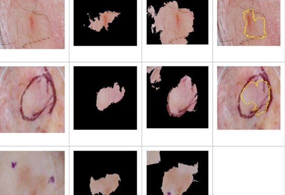
The researchers’ work focused on determining how the algorithm “thinks”, thus developing an “explainer”. This highlights on the photographs the areas on which the system focuses to detect melanoma. Visually, it can be seen how different pixels are illuminated in yellow. Thus, experts can know which area the “black box” algorithm is “looking at” and what logical process it follows to determine whether, for example, a spot is a melanoma or not.
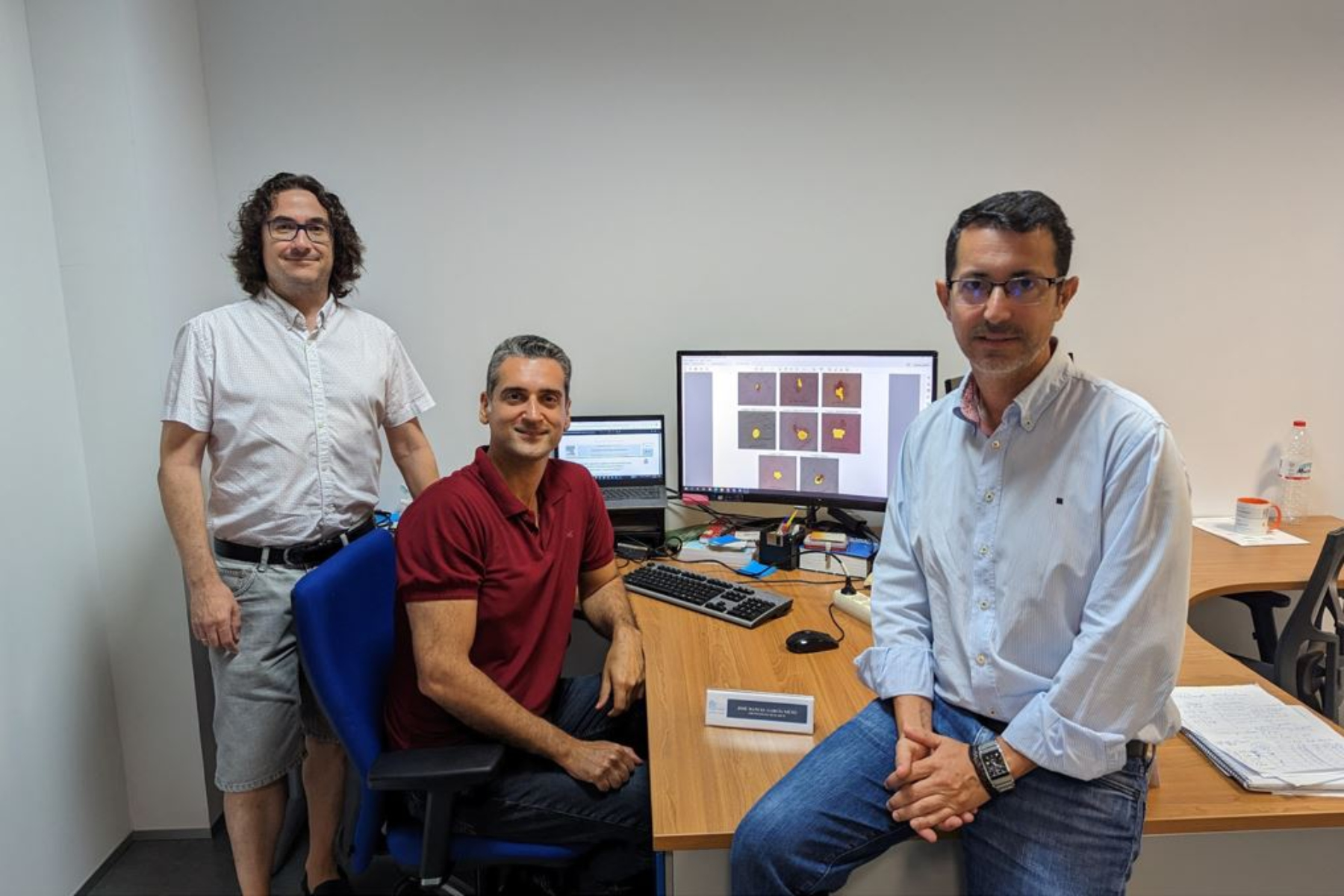
To test the effectiveness of this “explainer,” the Khaos Research Group at the Institute of Software Technology and Engineering, has assembled a set of images, drawn from free banks and privately held medical databases, containing spots, moles, freckles and other skin markings. Typically, doctors look at changes in shape, color, size or texture to determine whether they are benign or have progressed to melanoma. Thus, they have noticed that when the algorithm gets it right, it looks at very characteristic areas of skin tags; like physicians, while when it misses, it looks at less representative areas. “It detects different skin lesions such as melanoma, nevus, keratosis and healthy moles,” says University of Malaga researcher José Manuel García-Nieto.
To test the effectiveness of this tool, the experts have counted on the collaboration of Dr. Miguel Ángel Berciano (Intercenter Oncology Unit of the Virgen de la Victoria University Hospital), who, together with his collaborators, has checked the algorithm’s responses and has guided the researchers on the veracity of their answers and on the areas of interest for detecting possible pathologies in the skin marks.
Thus, the researchers have verified that the “explainer” works and that, based on it, the algorithm can be “taught” to follow the appropriate steps to detect the most complicated cases of melanoma. “The program does not replace in any case the work and diagnosis of a physician, but we propose it as a tool for pre-diagnosis of melanoma, as is already done in other hospitals with artificial intelligence that detects breast cancer,” concludes García-Nieto.
This work, which has been financed by the Ministry of Science and Innovation and the University of Malaga’s own funds, has as its next step the transfer of this “explainer” to domains other than the medical field, such as agriculture or environmental observation of the Earth.

